

1. Introduction
CT image denoising is one of the crucial parts of CT imaging and is widely utilized in medical science in recent years. During the generation of CT images, noise can be introduced through various ways such as the uncertainties in the physical measurements, position and operation of the equipment, and so on. The denoising of CT images can be divided into three domains: (1) Spatial Domain, (2) Transform Domain, and (3) Hybrid methods. Spatial domain consists of filters that are applied directly on the CT image to remove or reduce the noise. They can be linear and non-linear filters, anisotropic diffusion, total variation and dictionary learning methods, and Non-local means filters. In transform domain the input image or data is decomposed into a scale-space representation for image denoising. It includes wavelet based denoising, thresholding methods, shrinkage rules and so on. Finally, the hybrid methods consist of combinations of methods from both spatial and transform domain to yield better denoising results. Wiener filter is a filter in wavelet transformation that measures Mean Square Error (MSE) to give satisfactory denoised images. Thresholding is a process that not only suppresses the noise from the CT images but also helps in preserving the edges and corners of the image and hence the condemnatory and critical information in the image is preserved.
Rest of paper is organized as: section 2: a brief literature survey, Objectives, Methodology used, Merits and Demerits; Section 3: a comparative study; and lastly conclusion is drawn in section 4.
2. DWT based thresholding
The filters that make up the spatial domain are applied directly to the CT image in order to get rid of or significantly reduce the noise. Non-local means filters, linear and non-linear filters, anisotropic diffusion methods, total variation learning methods, and dictionary learning methods are some examples of these types of filters. In the transform domain, the process of image denoising commences with the input image or data being decomposed into a scale-space representation. It features thresholding methods, shrinkage rules, and denoising based on wavelets, among other things. In conclusion, the hybrid approaches are combinations of methods that come from the spatial domain as well as the transform domain. These methods produce superior denoising outcomes. DWT is one of the effective tools of image processing where image processing-based applications are utilized by getting approximation and detail parts of DWT. In image denoising, thresholding is applied on detail parts. After that inverse DWT can be performed to get denoised images as shown in Fig 1.
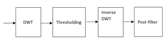
Figure 1. Wavelet based image denoising.
3. Literature survey: objectives, methodology used, merits and demerits
In the transform domain, the process of eliminating noise from an image begins with a representation of the image or data in scale space. It includes, among other things, methods for defining thresholds, rules for shrinking, and a method for removing noise based on wavelets. A summary of CT image denoising is shown in Table 1.
Table 1. A review on CT imae denoising.
Papers | Objective | Advantages | Disadvantages |
Goldstein et al, 2009 [1]. | This study aims to address multiple issues contingent to L-1 regularization, by the application of concepts of compressed sensing along with Rudin–Osher–Fatemi. | Application of Gauss–Seidel and Fourier transform makes it parallel in nature and the method shows seminal efficacy within limited time-space complexity as compared to fellow second order methods. | The absence any comprehensive literature review or competitive analysis for qualitative – quantitative analysis limits the scope of the study, making it appear parochial in nature. |
Elad et al, 2006 [2]. | Removal of zero-mean white and homogeneous Gaussian additive noise is performed by implementing optimized K-SVD algorithm. | This study successfully solves the issue of limited image patches when using K-SVD algorithm by early definition of global image. The approach of deliberating on HD image data set works in favour. | A detailed and contemporary literature survey or comparative analysis would edify the significant of the research and increase its scope for future studies in the purview. |
Mustafa et al, 2011 [3]. | The concept of bilateral filtering is extended to propose novel multi resolution bilateral filter (MRBF) with wavelet sub band, implementing concept of method noise. | This study understands the paucity of methods that are robust on real image data while also preventing artefact generation while noise suppression under limited space-time complexity. | A detailed and contemporary literature survey or comparative analysis would edify the significant of the research and increase its scope for future studies in the purview. |
Borsdorf et al, 2008 [4] | CT image denoising method is proposed under the assumption that unrelated noise resurfaces post reconstruction such that structure is preserved and noise does not return post reconstruction from multiple slices of data. | The method proposed addresses the issue of image structure preservation and resurfacing of noise post reconstruction. Adaptive and easy to implement nature of method provides it an advantage over others. | The quantitative and qualitative comparisons of the proposed method is limited and parochial. Absence of comparative analysis limits the scope of presented method and questions it’s efficacy. |
Rabbani et al, 2009 [5] | Propose a spatially adaptive medical image denoising method for removal of additive Gaussian or Rayleigh noise by the identification of heavy tail distribution in the noise pattern. | Parameters are deliberated using local information making proposed method to be spatially adaptive. Due to the application of accurate statistical models, artefacts introduced are not visible in the output image. Comparative analysis is detailed and cogent | The sequential multistep nature of the method proposed makes it complex to understand and cumbersome to implement. The dataset used is limited and stimulated, limiting method’s scope on variegated medical images. |
Blu et al, 2007 [6] | State of art image denoising technique is presented by implementing the concept of image-domain minimization to handle extra or undecided wavelet transforms as well. | The proposed method addresses the issue of noise creation by undecided wavelet transforms and solves the same by implementing the concept of image domain minimization. The results obtained are cogent and complete. | A detailed and contemporary literature survey or comparative analysis would edify the significant of the research and increase its scope for future studies in the purview. |
Chen et al, 2020 [7]. | Low CT and medical image denoising is performed by proposing Fractional-order Residual Convolutional Neural Network (FRCNN) for Low- Dose CT (LDCT) | Addition of skip connections successfully optimize the Convolutional neural network and this method performs low dose CTs denoising with image detail preservation, also solving health hazards related to high dose CT scans. | The multistep nature and extensive requirement of time-space complexity makes it cumbersome to implement as a unified framework. Also, a detailed quantitative analysis could edify its significance. |
Trung et al, 2020 [8]. | Low dose CT image denoising approach is proposed by implementing the concept of path based image denoising via splitting image into bands of low, medium and high frequency bands. | The method provides the claimed low dose CT image denoising method with almost complete detail preservation while proving its robustness with results on stimulated and real noisy CT and medical images. | The method loses its efficacy in case of image decomposition which can be improved. The splitting of images and patch wise denoising is a lengthy and time consuming process. |
Li et al, 2020 [9] | Cyclic generative adversarial network or CyclicGan is implemented on low dose CT scan images for denoising and detail preservation via image translation from low dose CT images to high dose CT images. | The comparative analysis performed is complete and cogent, and the literature compared also presents a detailed view of the proposed methodology. The method fulfils its claim of image detail preservation. | The method presented performs image translation via a primordial method that may or may not work on variegated types of real noisy image. |
Kumar et al, 2013 [10]. | The proposes a novel image denoising method that addresses the issue of loss of image details, absence of robustness, loss of image resolution and disorientation of pixels when denoised. | This successfully fulfils the claims of proposing a noise suppression method that preserves almost complete image details, maintains image resolution and acts robustly, which classical NLM filters lack. | The study is comprehensive and complete of current scenario but it fails to elaborate upon future scope or its application on variegated types of real noisy images. |
Diwakar et al, 2020 [11]. | Low dose CT image denoising the performed by implementing concepts of NLM, method noise and wavelet thresholding. | The results obtained show seminal numbers for all the deliberated matrices of qualitative analysis. The research study and its numerous sections are complete and concise. | A comprehensive and detailed comparative analysis of contemporary studies could further edify the significance of the study. |
Diwakar et al, 2022 [12] | State of art low dose COVID CT scan or medical image denoising approach is proposed for real noisy CT scan images. | The method performs low dose real COVID CT images noise suppression with detail preservation, image quality preservation, and preventing artefact generation in output image. The comparative analysis and literature is complete and cogent. | The study is comprehensive and complete of current scenario but it fails to elaborate upon future scope or its application on variegated types of real noisy images. |
4. Results and discussion
The outcome images are compared with certain already developed approaches in order to check the outcomes of the works being suggested. Methods used for contrast include Dictionary Learning and K-SVD-based denoising, Total Variability, NLM filter-based picture denoising and its noise thresholding method (NLFMT), SURELET-dependent de-noising, WBBT (Wavelet Based Bivariate Thresholding), wavelet based de-noising using correlation analysis, (WBCA), multiresolution Bilateral filter.
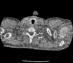
(a) (b) (c) (d)
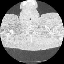
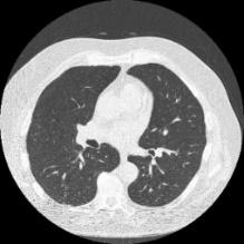
Figure 2. CT image dataset.
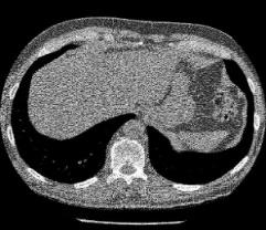
Fig 2 shows the CT image dataset, Fig 3: shows the outcome of total Variation, Fig 4: shows the outcome of Dictionary Learning and K-SVD-based denoising, Fig 5: shows the outcome of NLFMT filters. Fig 6: indicates the effect of denoising dependent on SURELET. It has been found that sharp picture information will get lost as the noise increases. The effects of wavelet-based denoising using correlation analysis, wavelet-based denoising using multi-resolution Bilateral filter, SURELET and wavelet-based bivariate thresholding in homogeneous regions are satisfactory however sharpness is not upto the mark. SURELET and multi-resolution dependent filtering do not produce acceptable outcomes and struggle with increasing noise level. Wavelet based bivariate thresholding method and wavelet based denoising using correlation analysis fails as the noise level in the image increases.
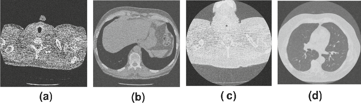
Figure 3. Outcomes of [2].
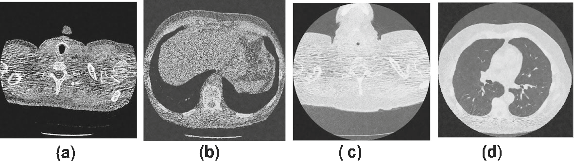
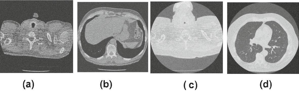 Figure 4. Outcomes of [3].
Figure 5. Outcomes of [4].
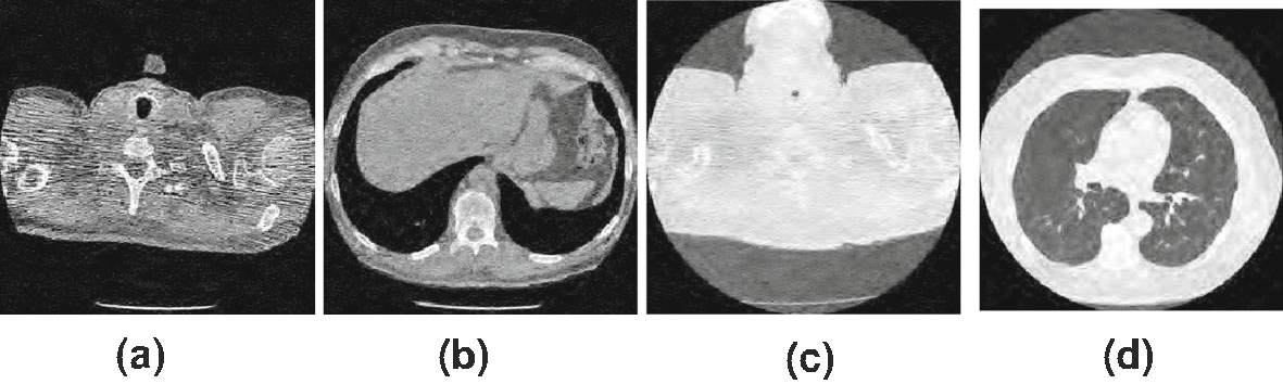
Figure 6. Outcomes of [5].
The already existing methods and the proposed framework are set side by side with Structure Similarity (SSIM) as shown in table 2.
Table 2. SSIM of noiseless CT images.
Input | CT Image Dataset 1 (Average results on 80 images) | CT Image Dataset 2 (Average results on 70 images) | ||||||
Σ | 10 | 20 | 30 | 40 | 10 | 20 | 30 | 40 |
[1] | 0.7821 | 0.7993 | 0.7078 | 0.6593 | 0.8362 | 0.7953 | 0.7609 | 0.6481 |
[2] | 0.7738 | 0.7964 | 0.7076 | 0.6577 | 0.8307 | 0.7898 | 0.7571 | 0.6561 |
[3] | 0.7564 | 0.7872 | 0.7024 | 0.6551 | 0.8208 | 0.7777 | 0.7451 | 0.6413 |
[4] | 0.7428 | 0.7777 | 0.6984 | 0.6518 | 0.8122 | 0.7703 | 0.7394 | 0.6306 |
[5] | 0.7955 | 0.8159 | 0.7134 | 0.6635 | 0.8451 | 0.7026 | 0.7681 | 0.6556 |
5. Conclusion and future scope
There are many dimensions to which this paper can be extended due to the adaptive nature of the denoising methods. The more intelligent use of method noise can improve the results. The paper uses a bilateral filter in the proposed method that processes the approximate part. The approximate part holds the residual content. This content can be processed/ filtered using many other methods too. Those different filters can be explored as well. Apart from this, various internal parameters can be changed and checked as well.