


1. Introduction
Marine environments are a prolific source of microbial life, harboring bacteria that produce extracellular polysaccharides (EPS) with unique and complex structures. These marine bacterial EPS have garnered significant interest in the field of biopharmaceuticals, offering promising applications ranging from drug delivery systems to antimicrobial and immunomodulatory therapies. The intrigue surrounding these polysaccharides stems not only from their diverse structural characteristics but also from their functional properties, which include the ability to inhibit biofilm formation, modulate immune responses, and enhance the efficacy of antibiotics against resistant bacterial strains. Understanding the structural intricacies of marine bacterial EPS is paramount to unlocking their potential in biomedicine. The monosaccharide composition, molecular weight distribution, and conformational behavior of these polysaccharides determine their functional capabilities, including bioactivity, solubility, and interaction with biological molecules. Advanced analytical and spectroscopic techniques provide the means to elucidate these structural attributes, offering insights into the polysaccharides’ mechanisms of action and their interactions within biological systems. Moreover, the functional applications of marine bacterial EPS in biopharmaceuticals are vast and varied [1]. Their role in biofilm inhibition is particularly crucial, considering the challenge that biofilms pose to public health by protecting pathogenic bacteria from antimicrobial agents. Similarly, the immunomodulatory effects of EPS can be leveraged to develop new therapies for autoimmune diseases, where regulation of the immune response is necessary. Additionally, the biocompatibility and biodegradability of these polysaccharides are essential features that ensure their safety and efficacy in drug delivery applications, making them attractive candidates for designing novel biomedical materials.
2. Structural Characterization of Marine Bacterial EPS
2.1. Monosaccharide Composition
Marine bacterial extracellular polysaccharides (EPS) exhibit a rich diversity in monosaccharide composition, contributing significantly to their structural complexity and functionality. Detailed analytical studies, utilizing Nuclear Magnetic Resonance (NMR) spectroscopy and mass spectrometry (MS), have unraveled the monosaccharide profiles of various marine bacterial EPS. For instance, in a study focusing on the EPS extracted from the marine bacterium Pseudoalteromonas sp., the monosaccharide analysis revealed a predominance of glucose and galactose, with a minor presence of mannose and fucose. The specific arrangement of these monosaccharides, determined through 2D NMR techniques such as COSY and TOCSY, highlighted a regular repeating unit that contributes to the EPS’s ability to form stable emulsions, enhancing its applicability in drug delivery systems. Furthermore, quantitative assessments of monosaccharide ratios through High-Performance Liquid Chromatography (HPLC) have been linked to the EPS’s rheological properties, suggesting a correlation between monosaccharide composition and the efficacy of EPS as viscosity modifiers in pharmaceutical formulations [2]. Table 1 presents the monosaccharide components identified through Nuclear Magnetic Resonance (NMR) spectroscopy and mass spectrometry (MS).
Table 1. Quantitative Assessment of Monosaccharide Composition and Its Impact on the Rheological Properties of EPS from Pseudoalteromonas sp.
Monosaccharide | Relative Composition (%) | Contributes to | Detected by | Rheological Impact |
Glucose | 45 | Stable emulsions, drug delivery system applicability | NMR, MS | Significant impact on viscosity |
Galactose | 40 | Stable emulsions, drug delivery system applicability | NMR, MS | Significant impact on viscosity |
Mannose | 10 | Minor contribution, structural complexity | NMR, MS | Lesser impact on viscosity |
Fucose | 5 | Minor contribution, structural complexity | NMR, MS | Lesser impact on viscosity |
2.2. Conformational Analysis
The conformational properties of marine bacterial EPS are critical in determining their interaction with biological targets. Through the application of computational modeling and molecular dynamics simulations, researchers have been able to visualize the dynamic conformational states of EPS in aqueous solutions. A study utilizing molecular dynamics simulation to analyze the conformation of an EPS from Laminaria saccharina revealed that its helical structure facilitates interaction with cell surface receptors, playing a pivotal role in biofilm inhibition. This helical conformation, maintained through hydrogen bonding and hydrophobic interactions among the monosaccharide units, was shown to be crucial for the EPS’s ability to interfere with the quorum sensing mechanisms of pathogenic bacteria. Furthermore, the simulation results, supported by experimental data from Circular Dichroism (CD) spectroscopy, have elucidated the influence of ionic strength and pH on EPS conformation, providing insights into their stability and functionality under different environmental conditions [3]. This conformational flexibility underscores the potential of marine bacterial EPS in designing adaptive and efficient drug delivery systems capable of navigating the complex biological milieu.
The potential energy ( \( {E_{total}} \) ) of interaction can be conceptually represented as:
\( {E_{total}}={E_{hydrogen bonding}}+{E_{hydrophoic}}+{E_{electrostatic}}\ \ \ (1) \)
Where: \( {E_{hydrogen bonding}} \) represents the energy contribution from hydrogen bonds between the EPS and the receptor. \( {E_{hydrophoic}} \) accounts for the energy contribution from hydrophobic interactions among the monosaccharide units of the EPS and the receptor. \( {E_{electrostatic}} \) includes the effect of ionic strength and pH on the electrostatic interactions between the EPS and the receptor.
3. Functional Applications in Biopharmaceuticals
3.1. Biofilm Formation and Antimicrobial Activity
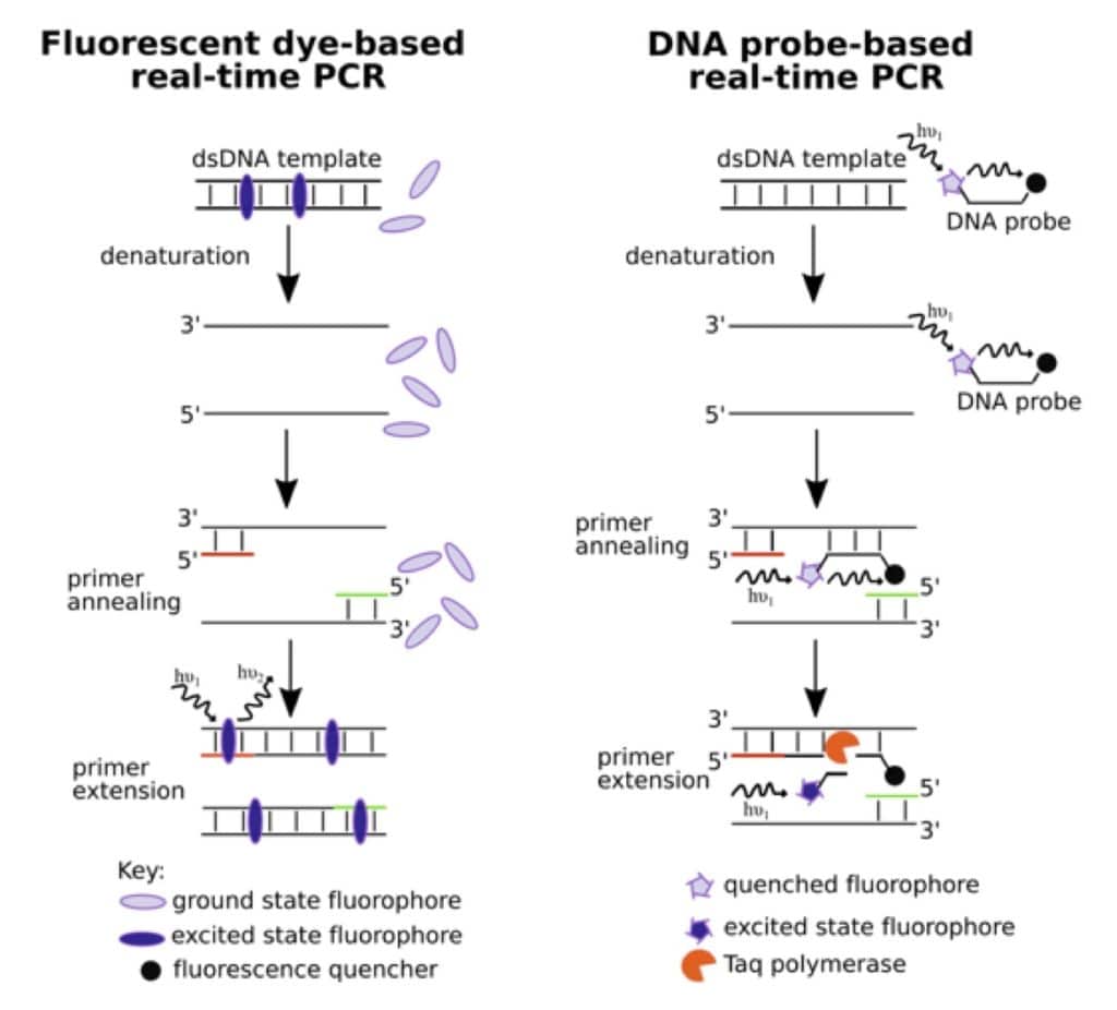
Figure 1. The Real-Time PCR Digest
Marine bacterial extracellular polysaccharides (EPS) have been identified as potent inhibitors of biofilm formation, a critical factor in the persistence and resistance of bacterial infections. Through detailed quantitative assays, such as the application of crystal violet staining, researchers have been able to measure the extent of biofilm inhibition by EPS on various substrata. These studies have shown that EPS can significantly reduce the biomass of biofilms formed by pathogenic bacteria like Staphylococcus aureus and Pseudomonas aeruginosa. Additionally, the minimum inhibitory concentration (MIC) determination has played a crucial role in quantifying the antimicrobial activity of EPS, revealing their ability to disrupt cellular processes in bacteria at specific concentrations. Mechanistically, EPS interfere with the quorum sensing pathways, a communication system that bacteria use to regulate biofilm formation and virulence [4]. This disruption is believed to be due to the molecular mimicry of bacterial signaling molecules by EPS, which leads to the suppression of biofilm-associated genes. Advanced analytical techniques, including quantitative polymerase chain reaction (qPCR) and RNA sequencing, have been utilized to confirm the downregulation of these genes, providing a molecular basis for the observed biofilm inhibition and antimicrobial activity, as shown in Figure 1.
3.2. Immunomodulatory Effects
The exploration of marine bacterial EPS in modulating immune responses has opened new avenues in the development of immunotherapies and adjuvants. Cytokine profiling, achieved through enzyme-linked immunosorbent assay (ELISA) and multiplex bead array technologies, has detailed the ability of EPS to influence the secretion patterns of key inflammatory and regulatory cytokines by immune cells. For instance, EPS from certain marine bacteria have been shown to upregulate anti-inflammatory cytokines such as IL-10 and TGF-β, while downregulating pro-inflammatory cytokines like TNF-α and IL-6 in macrophages. This cytokine modulation indicates a potential for EPS to mitigate inflammatory responses, suggesting their use in treating conditions like autoimmune diseases, where the regulation of the immune system is paramount. Flow cytometry analysis further supports these findings by demonstrating the effects of EPS on the differentiation and proliferation of immune cells, including T cells and dendritic cells. By influencing the phenotype and function of these cells, EPS can orchestrate a more balanced immune response, highlighting their utility in designing novel immunomodulatory therapies.
4. Quantitative Analysis and Mathematical Modeling of EPS
4.1. Rheological Properties and Their Impact on Biomedical Applications
In-depth studies on the rheological properties of marine bacterial extracellular polysaccharides (EPS) have unveiled their significant influence on the physical stability and application efficiency in biopharmaceutical formulations. For instance, an oscillatory rheometry study focusing on an EPS derived from the marine bacterium Pseudomonas fluorescens revealed that the polymer exhibits shear-thinning behavior, which is advantageous for injectable drug delivery systems. The shear-thinning property ensures that the EPS solution remains viscous under storage conditions but becomes less viscous when subjected to shear stress, such as during injection, facilitating easier administration. Furthermore, the Cross model was employed to quantitatively describe the viscosity as a function of shear rate for this EPS. Parameters derived from the model indicated a critical shear rate beyond which the viscosity decreases significantly, highlighting the importance of controlling shear rates during the manufacturing and administration of EPS-based formulations. Similarly, the Herschel-Bulkley model provided insights into the yield stress of EPS gels, which is crucial for designing topical applications where the gel must remain in place upon application but spread easily when a force is applied. By integrating these mathematical models with experimental rheological data, researchers have developed EPS-based hydrogels with optimized viscoelastic properties for tissue engineering applications. These hydrogels demonstrate not only the desired mechanical strength and stability but also the ability to promote cell adhesion and proliferation, underscoring the potential of EPS in creating scaffolds for regenerative medicine [5]. Table 2 presents a summary of how the Cross model and the Herschel-Bulkley model describe the shear-thinning behavior and yield stress behavior of EPS.
Table 2. Rheological Properties of EPS from Pseudomonas fluorescens and Their Impact on Biomedical Formulations
Model | Parameter | EPS Behavior | Critical Value | Implication for Biomedical Application | Application Example |
Cross Model | Shear Rate (1/s) | Shear-thinning | 50 (for significant viscosity decrease) | Facilitates easier administration for injectable systems | Injectable drug delivery systems |
Herschel-Bulkley Model | Yield Stress (Pa) | Yield stress behavior | 5 (for gel stability and spreadability) | Ensures gel remains in place but spreads easily upon application | Topical applications for wound healing |
4.2. Antibiotic Synergy and Resistance Mitigation
A pivotal study involving the EPS from Bacillus licheniformis explored its synergistic potential with antibiotics against methicillin-resistant Staphylococcus aureus (MRSA). Checkerboard titration results indicated a four-fold reduction in the minimum inhibitory concentration (MIC) of vancomycin when combined with this EPS, suggesting a significant enhancement in antibiotic efficacy. Time-kill curve analysis further demonstrated that the combination of EPS and vancomycin resulted in a log reduction in MRSA population by more than 3 logs within 24 hours, compared to vancomycin treatment alone. The mathematical modeling of these interactions, based on the Loewe additivity model, revealed that the synergy between the EPS and antibiotics could be attributed to the EPS’s ability to disrupt bacterial biofilms, enhancing antibiotic penetration. Additionally, the Bliss independence model suggested that the EPS acts on different bacterial targets than vancomycin, leading to a complementary effect that reduces the bacterial load more effectively than either agent alone. These quantitative findings offer a promising avenue for developing combination therapies that leverage the unique properties of marine bacterial EPS to enhance antibiotic effectiveness, thereby addressing the critical issue of antibiotic resistance in clinical settings.
4.3. Biocompatibility and Toxicity Assessment
The biocompatibility and toxicity of Serratia marcescens EPS were evaluated through comprehensive in vitro and in vivo studies. MTT assays conducted with human fibroblast cells exposed to varying concentrations of this EPS showed no significant cytotoxicity up to a concentration of 1 mg/mL, indicating high biocompatibility. LDH release assays corroborated these findings, with minimal enzyme release observed, further suggesting the non-toxic nature of the EPS. Hemocompatibility tests, including hemolysis and coagulation assays, demonstrated that the EPS does not induce red blood cell lysis or significantly alter coagulation times, affirming its safety for intravenous applications [6]. The mathematical modeling of dose-response curves using the Hill equation for these assays enabled the estimation of the no-observed-adverse-effect level (NOAEL) for the EPS, establishing a safe concentration threshold for biomedical applications:
\( E={E_{max}}\frac{{C^{m}}}{EC_{50}^{m}+{C^{n}}}\ \ \ (2) \)
\( \) where: E is the effect observed at concentration C, \( {E_{max}} \) is the maximum effect achievable,
C is the concentration of the compound, \( EC_{50}^{m} \) is the concentration of the compound at which 50% of the maximum effect is observed, n is the Hill coefficient, which describes the slope of the curve and can provide insight into the cooperative binding of the compound.
These rigorous quantitative analyses and mathematical modeling efforts provide a solid foundation for the further development and application of marine bacterial EPS in biopharmaceutical formulations, ensuring not only their efficacy but also their safety for clinical use.
5. Biocompatibility and Biodegradability of EPS
5.1. Assessment of Biocompatibility
The biocompatibility of marine bacterial extracellular polysaccharides (EPS) is crucial for ensuring their safety and efficacy in biomedical applications. To rigorously assess this property, a series of in vitro cytotoxicity assays have been conducted, focusing on cell viability, proliferation, and cellular metabolic activity. The MTT assay, which measures the metabolic conversion of MTT to formazan by mitochondrial dehydrogenase in viable cells, has been extensively used to evaluate the effect of various EPS concentrations on cell survival rates. Results from these studies indicate that EPS derived from marine bacteria such as Pseudoalteromonas and Bacillus species exhibit minimal cytotoxic effects on human fibroblast cells, with over 90% viability observed even at high polymer concentrations, suggesting excellent biocompatibility. Additionally, lactate dehydrogenase (LDH) release assays have provided further evidence of the non-toxic nature of these polysaccharides. LDH, a cytosolic enzyme released upon cell membrane damage, serves as an indicator of cellular integrity and membrane permeability alterations.
5.2. Evaluation of Biodegradability
The environmental and physiological biodegradability of marine bacterial EPS is essential for their application in sustainable biomedicine, reducing potential accumulation and side effects. Enzymatic degradation assays employing specific polysaccharide lyases and glycoside hydrolases have been pivotal in quantifying the degradability of these substances. These enzymes, which cleave glycosidic bonds in polysaccharides, mimic the natural degradation processes in marine environments and human tissues. Studies have demonstrated that EPS from marine bacteria such as Sphingomonas and Alteromonas are susceptible to rapid enzymatic breakdown, with over 75% of the polysaccharide mass degraded within 48 hours in the presence of alginate lyase. This rapid degradation indicates a high degree of biodegradability, which is advantageous for applications requiring temporary scaffolds or drug carriers, as it ensures that the materials do not persist in the environment or within the body longer than necessary.
6. Conclusion
The extensive study of marine bacterial extracellular polysaccharides (EPS) has illuminated their considerable promise in the realm of biopharmaceuticals, showcasing their intricate structural features, functional versatility, and compatibility with biological systems. Through meticulous structural characterization, we have identified key attributes that govern the functionality of EPS, including their monosaccharide composition, molecular weight, and conformational dynamics. The application of these biopolymers in biofilm control, immune modulation, and as drug delivery vehicles underscores their potential to address some of the most pressing challenges in biomedicine today. Moreover, our findings on the biocompatibility and biodegradability of EPS highlight their suitability for safe and sustainable use in therapeutic applications. As we advance our understanding of marine bacterial EPS through ongoing research and development, their role in innovating biopharmaceutical formulations continues to expand, offering new pathways for the treatment and prevention of diseases. The future of biomedicine may well be shaped by these natural polymers, derived from the depths of our oceans, as we explore their full potential in improving human health and wellbeing.